


Mast Cell Tumors in Dogs
Background
Mast cells are a type of white blood cell that play a key role in a dog’s immune system. They contain granules that release histamine and other chemicals involved in allergic reactions and fighting off infectious agents like parasites and bacteria. When mast cells become cancerous they form a mass called a mast cell tumor (MCT).
Mast cell tumors are the most common malignant skin tumor found in dogs. They may be seen in dogs of any age but occur most commonly in dogs who are 8 to 10 years old. They can develop on any part of a dog’s body, including internal organs, but the limbs, lower abdomen, and chest are the most common sites. If these tumors spread, they typically spread first lymph nodes near the tumor followed by the liver and spleen.
Risk Factors
Any dog can develop a mast cell tumor, but the following breeds are at a greater risk:
- American Staffordshire terriers
- Boxers
- French bulldogs
- Golden retrievers.
- Labrador retrievers
- Pugs
- Rhodesian ridgebacks
- Shar-peis
Signs
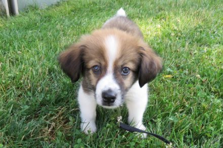
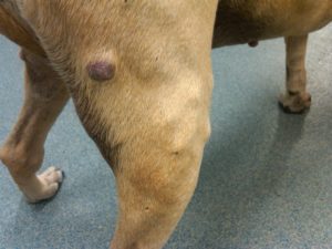
Mast cell tumors vary in appearance. Some look like raised, hairless bumps on or just below the surface of the skin. Others appear as red, ulcerated, bleeding, bruised, or swollen growths. Some tumors stay the same size for months or years, some may spontaneously grow and shrink, while others grow rapidly over days or weeks. Additional signs such as vomiting, loss of appetite, and dark tarry stool typical of intestinal bleeding are also possible.
Diagnosis
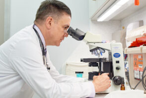
Most mast cell tumors can be diagnosed with a simple needle aspirate of the tumor. This quick procedure is done while the dog is awake, and involves inserting a thin, hollow needle into the mass to collect a sampling of cells, which are then examined under a microscope.
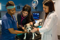
Treatment
Surgical removal of a mast cell tumor is often the initial mainstay of treatment. The best chance of removing the entire tumor is to remove a 1 ¼ – inch margin in all directions around the growth. This results in a larger incision than most pet owners expect.
After the mast cell tumor is surgically removed, the sample will be sent to a veterinary pathologist who will look at the tumor using a microscope and make a variety of assessments about the tumor. The pathologist will record how abnormal the cells look, how much the cells invade into the normal tissue, how many cells are dividing, and what the center or nucleus of the cells look like. The pathologist then “grades” the tumor by assigning it a number: 1, 2, or 3. The more abnormal the cells, the higher the grade. The lower the grade, the more favorable the prognosis. If a biopsy indicates the tumor has not been completely removed or if the tumor cannot be removed, or if there is more than one tumor, veterinary oncologists may recommend radiation therapy or chemotherapy to try to control the mast cell tumor.
Radiation therapy is typically used for mast cell tumors that have not spread or that have only spread to a nearby lymph node. For some mast cells tumors, radiation can provide years of cancer-free time. In general, most dogs do well with radiation.
Chemotherapy is typically used for mast cell tumors that are grade 2 or 3 or that have spread elsewhere in the body. Chemotherapy may be given as a pill at home by the pet parent or as an injection given by the medical oncology team as an outpatient appointment at AMC. In general, most dogs do well with chemotherapy.
Prevention
Dogs who have had one mast cell tumors are at greater risk for developing additional tumors. Early detection and removing tumors when they are small and have not spread increases the likelihood of treatment success. For that reason, it is important to check your dog regularly for lumps and agree to a fine needle aspirate If your veterinarian suggests one for your dog.
Make an Appointment
Oncology
AMC's Cancer Institute is a revolutionary medical center, created to provide integrative and comprehensive care for pets diagnosed with cancer.
Learn More